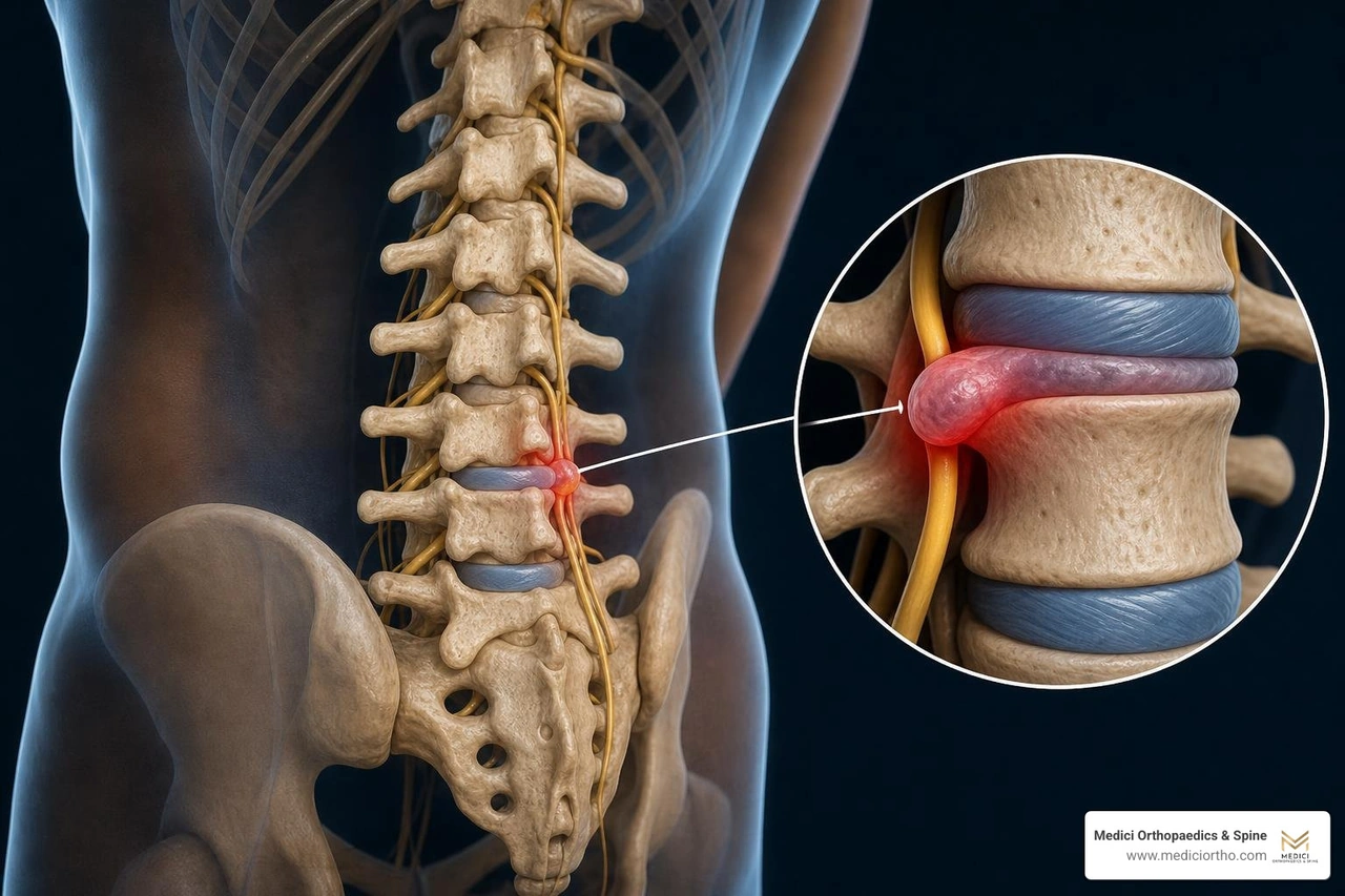
Lumbar disk compression affects millions of people — and it is one of the most common reasons adults seek medical care for back and leg pain. In fact, roughly 80% of people will experience at least one episode of low back pain in their lifetime.
Here is what lumbar disk compression means in plain terms:
| Question | Quick Answer |
|---|---|
| What is it? | A spinal disc in the lower back presses on a nearby nerve root or the spinal cord |
| Where does it happen? | 95% of cases occur at L4-L5 or L5-S1 |
| What does it feel like? | Lower back pain, leg pain (sciatica), numbness, tingling, or weakness |
| Does it heal on its own? | Yes — 85–90% of cases improve within 6–12 weeks without surgery |
| When is it an emergency? | If you lose bladder or bowel control, seek care immediately |
The pain can range from a dull ache in your lower back to a sharp, burning sensation that shoots down your leg. Some people feel it only when sitting. Others feel it constantly. The experience varies depending on which nerve is compressed and how much pressure is on it.
What makes this condition especially frustrating is that it often strikes active, otherwise healthy adults — people who want to keep moving, stay off medications, and avoid surgery if at all possible.
The good news? Most people do not need surgery. And understanding what is actually happening inside your spine is the first step toward getting the right care.
I'm Dr. Sonny Dosanjh, M.D., founder of Medici Orthopaedics & Spine, board certified in Physical Medicine & Rehabilitation, and fellowship trained at Emory University in Multidisciplinary Pain Management — and lumbar disk compression is one of the conditions I treat most frequently across our Metro Atlanta clinics. In this guide, I'll walk you through everything you need to know, from anatomy and diagnosis to the full range of treatment options available today.
To understand why your back or leg hurts, we must first look at the anatomy of the spine. The spine is a marvel of biological engineering made up of 33 vertebrae. In the lower back—the lumbar spine—we have 5 robust bony segments designed to carry the weight of your entire upper body.
Between these bones sit your intervertebral discs, which act as the spine's natural shock absorbers. Each disc consists of two main parts:
When these discs lose their structural integrity, they can compress nearby neural structures. However, patients often use terms like "herniated disc," "degenerative disc disease," and "spinal cord compression" interchangeably. In the medical community, we distinguish between these conditions because they require very different treatment pathways.
| Condition | Primary Pathology | Typical Symptoms | Primary Diagnostic Tool |
|---|---|---|---|
| Lumbar Disc Herniation | Soft inner nucleus tears through the tough outer ring, directly pinching a nerve root. | Sharp, shooting leg pain (sciatica), numbness, localized weakness. | MRI |
| Degenerative Disc Disease (DDD) | Age-related wear and tear where the disc loses water content (desiccation) and flattens. | Dull, chronic ache in the lower back that worsens with sitting or bending. | X-ray or MRI |
| Spinal Cord Compression | Pressure on the actual spinal cord (or the cauda equina in the lower lumbar spine). | Bilateral weakness, loss of coordination, bowel/bladder dysfunction (emergency). | MRI with contrast |
It helps to think of the difference between a bulging disc and a herniated disc like a jelly donut.
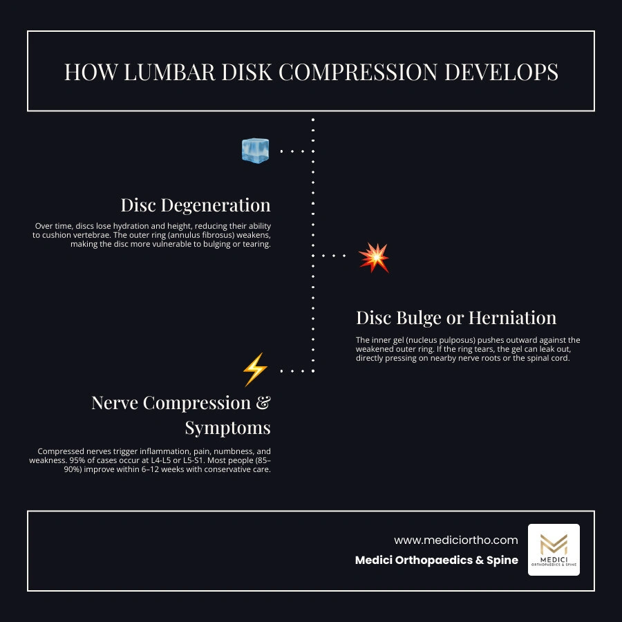
A Bulging Disc is like pressing down gently on the donut. The outer wall remains intact, but the entire disc bulges outward, encroaching on the surrounding space. The inner "jelly" has not broken through.
A Herniated Discs occurs when there is an actual tear in the tough outer ring (the annulus fibrosus). This allows the soft, jelly-like nucleus pulposus to squeeze out. This protruding material does not just mechanically pinch the nerve; it also releases inflammatory chemical agents that irritate the nerve root, causing intense, radiating pain.
As we age, our discs undergo a natural process called desiccation, meaning they gradually lose their water content and dry out. This is the hallmark of Degenerative Disc Disease, a condition that is heavily influenced by genetics and lifestyle factors.
As the discs flatten, the space between your vertebrae shrinks. To compensate for this structural instability, your body tries to stabilize the area by growing extra bone. These are known as bone spurs (osteophytes). Over time, the combination of flattened discs and bone spurs can narrow the open spaces within your spine—a condition called Spinal Stenosis. This narrowing squeezes the nerves traveling through the spinal canal, often causing pain or cramping in both legs when you stand or walk for long periods.
It is also vital to distinguish between a pinched individual nerve root (radiculopathy) and compression of the spinal cord itself (myelopathy).
The spinal cord actually ends around the L1-L2 vertebral level, just below the ribs. From that point downward, the spinal canal is filled with a bundle of nerve roots called the cauda equina (Latin for "horse's tail").
According to clinical resources like Spinal Cord Compression - StatPearls - NCBI Bookshelf , compression can be caused by extramedullary lesions (outside the spinal cord, such as herniated discs or bone spurs) or intramedullary lesions (inside the cord itself). Identifying the exact location and nature of the compression is critical for preventing long-term damage.
Why do these natural shock absorbers fail in the first place? In most cases, lumbar disk compression is not the result of a single, dramatic injury. Instead, it is the product of long-term wear and tear combined with specific risk factors.
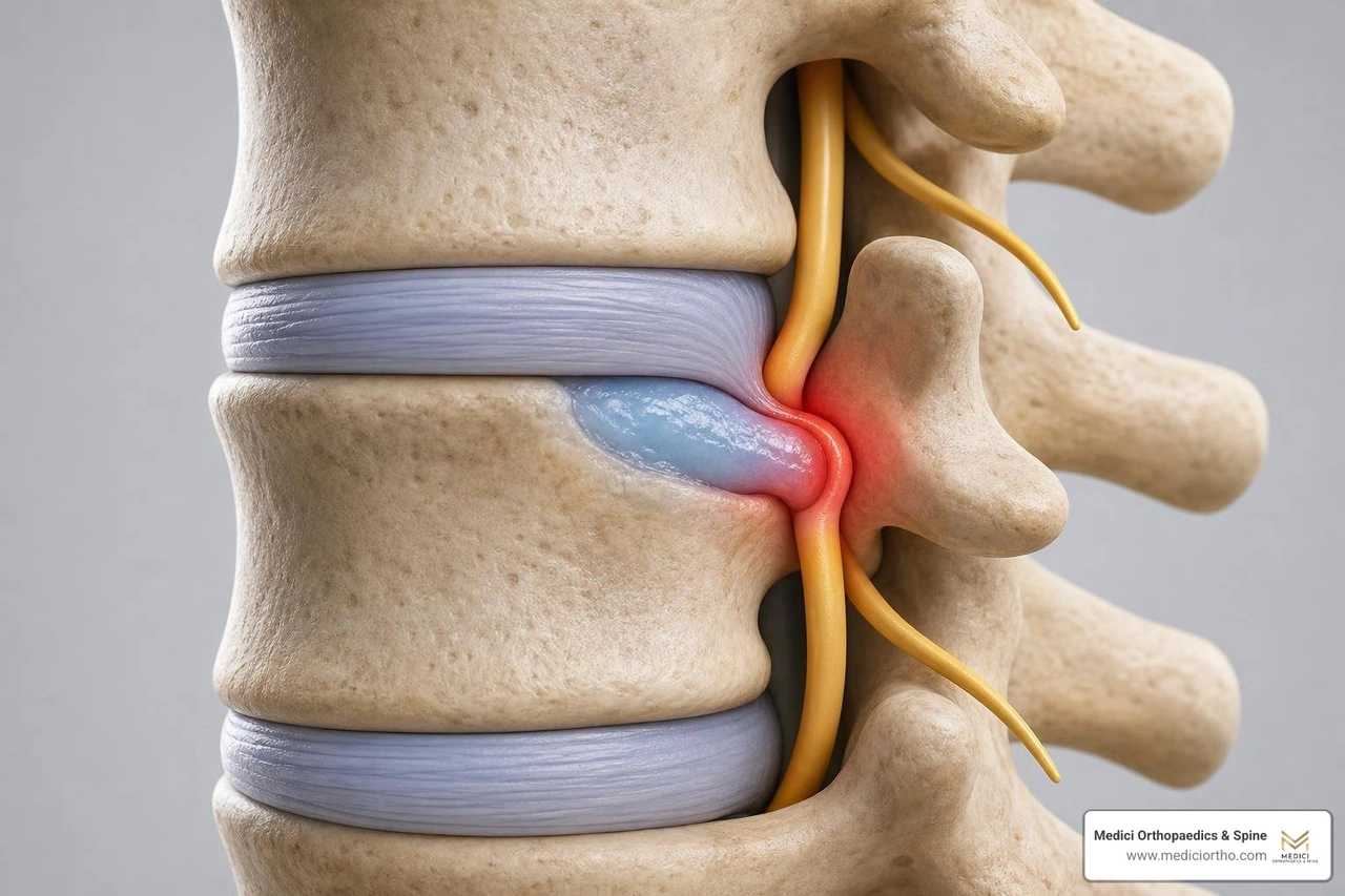
The primary mechanical driver is axial overloading—putting too much downward, vertical pressure on the spine. When you combine heavy lifting with twisting or bending, you create a perfect storm of mechanical forces that can tear the annulus fibrosus.
However, we now know that genetics play an even larger role than occupational hazards. If your parents or siblings struggled with herniated discs, your spinal structures may naturally be more prone to early wear and tear.
Several everyday lifestyle choices and physical factors can significantly accelerate disc degeneration:
At Medici Orthopaedics & Spine, we evaluate the entire kinetic chain rather than treating the lower back in isolation. A perfect example of this is "Knee-Spine Syndrome."
A fascinating 3D musculoskeletal analysis published in The Effect of Knee Extension Limitation on Lumbar Intervertebral Disc Compression Force During Walking: A 3D Musculoskeletal Analysis demonstrates how a limitation in knee extension (an inability to fully straighten your leg by 10 degrees or more) directly impacts your spine.
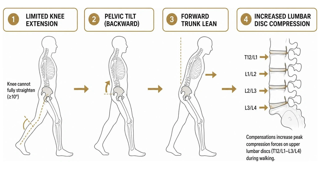
When your knee cannot fully straighten, your body compensates during walking by tilting your pelvis backward and leaning your trunk forward. This compensatory lean dramatically increases the peak compression forces acting on your upper lumbar intervertebral discs (T12/L1 down to L3/L4) with every single step you take. By restoring full range of motion to your knees through targeted physical therapy, we can actively reduce the mechanical overload on your lower back.
The symptoms of lumbar disk compression are rarely confined to the lower back itself. Because the nerves in your lumbar spine travel all the way down to your toes, a pinch in your back can feel like a fire in your foot.
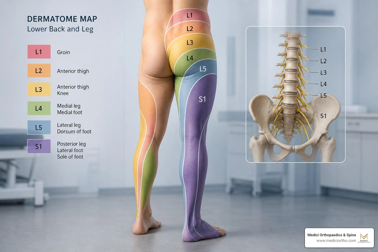
This radiating leg pain is commonly known as Sciatica. Depending on which nerve is compressed, you may also experience muscle spasms, localized numbness, or a loss of specific reflexes.
During a physical examination at our clinics in Snellville, Kennesaw, Marietta, or Atlanta, we use "dermatomal mapping" to pinpoint exactly which nerve root is under pressure. As outlined in the Lumbar Disc Herniation - Rheumatology and Orthopedics - Merck Manual Professional Edition , each lumbar nerve root corresponds to a specific area of sensation and motor control:
While most cases of lumbar nerve compression can be managed safely over time, there are certain "red flag" symptoms that represent a true medical emergency.
If a massive disc herniation completely blocks the spinal canal and compresses the cauda equina, it can cause irreversible nerve damage if not treated immediately. According to the Spinal Cord Compression - Neurology - MSD Manual Professional Edition , you must seek immediate emergency medical care if you experience:
A successful recovery starts with an accurate diagnosis. We do not just treat what we see on an imaging scan; we carefully correlate your physical symptoms with your diagnostic images to build a personalized treatment plan.
Before ordering expensive scans, we perform a series of physical tests designed to put gentle tension on your spinal nerves. If a nerve is compressed and inflamed, these movements will recreate your typical pain:
We strictly follow evidence-based guidelines when it comes to diagnostic imaging. As emphasized in Lumbar Disc Herniation - StatPearls - NCBI Bookshelf , we generally avoid ordering advanced imaging like an MRI during the first 6 weeks of acute back pain unless "red flags" are present.
Why? Because up to 90% of acute disc herniations resolve on their own within 6 to 12 weeks, and we want to avoid unnecessary medical costs and anxiety for our patients.
However, if your symptoms persist past 6 weeks despite conservative care, or if you have progressive neurological weakness, an MRI is the gold standard. With a diagnostic accuracy of approximately 97%, an MRI allows us to clearly see the soft tissues, the exact size of the disc herniation, and the degree of nerve root compression.
At Medici Orthopaedics & Spine, our philosophy is simple: we believe in delivering the most effective, least invasive, and least drug-dependent treatments medically available. We find that many of our therapies work synergistically—meaning they produce far better results when used together than when used in isolation.
For the vast majority of our patients in Metro Atlanta, we can achieve complete pain relief and restore your quality of life without surgery. Our comprehensive, non-surgical toolkit includes:
While we exhaust every non-surgical option first, surgery is sometimes the safest and most effective path forward. Surgical intervention is typically indicated if you experience progressive neurological deficits (like worsening foot drop), signs of cauda equina syndrome, or intractable pain that makes daily life impossible despite months of dedicated conservative care.
When surgery is necessary, we favor ultra-minimally invasive techniques. An Endoscopic Discectomy or microscopic discectomy allows a surgeon to remove only the tiny fragment of herniated disc that is pressing on the nerve through an incision no larger than a postage stamp. This minimizes damage to surrounding muscles, preserves spinal stability, and allows for a much faster, less painful recovery.
Yes, they absolutely can! It sounds surprising, but a herniated disc will naturally dry out (desiccate) and shrink over time. Through a natural process called phagocytosis, your body's immune cells recognize the displaced disc material as foreign and gradually break it down and reabsorb it.
This natural resorption typically occurs over a 6 to 9-month timeline. This is why we prioritize conservative management first; if we can control your pain and keep you active, your body will often do the heavy lifting of healing itself.
Sitting is often the enemy of a healing lower back. When you sit, you lose the natural forward curve (lordosis) of your lumbar spine, which shifts your body weight directly onto the discs.
In fact, clinical measurements show that sitting increases the mechanical pressure on your lumbar discs by approximately 40% compared to standing. If you already have a compressed nerve, this increased pressure can instantly trigger or worsen your sciatica. We highly recommend using an ergonomic lumbar support, taking standing breaks every 30 minutes, or using a height-adjustable standing desk.
Ignoring severe or progressive nerve compression can lead to permanent, irreversible damage. Over time, constant pressure starves the nerve of oxygenated blood, which can lead to:
Living with lumbar disk compression can feel overwhelming, but you do not have to navigate this journey alone. Whether you are dealing with a fresh flare-up of sciatica or chronic back pain that has kept you on the sidelines for years, there is hope for a full recovery.
At Medici Orthopaedics & Spine, led by Dr. Sonny Dosanjh, M.D., we are committed to helping you find lasting relief using the most advanced, non-surgical, and minimally invasive treatments available. We don't just treat your symptoms—we look at your entire body, from your knees to your spine, to find and fix the root cause of your pain.
If you are ready to take control of your spinal health and optimally restore your quality of life, we invite you to take the next step. Learn more about how we diagnose and treat Lumbar Radiculopathy (Pinched Nerve) and schedule your comprehensive evaluation at our clinics in Atlanta, Snellville, Kennesaw, or Marietta. Let us help you get back to living life without limits.
At Medici, you’re more than your MRI.
We take time to hear your story, understand your pain, and create a plan that actually works for you.

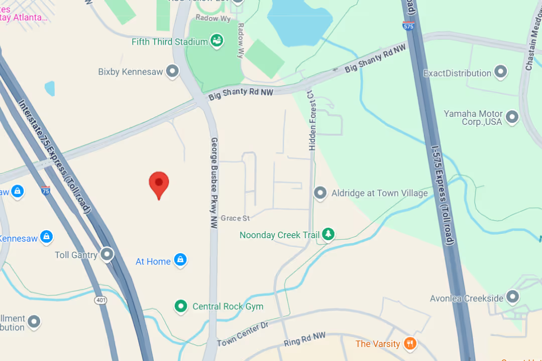
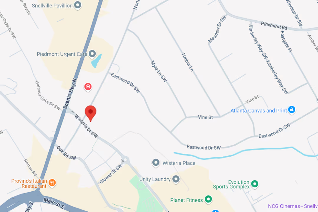
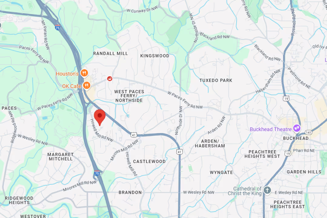
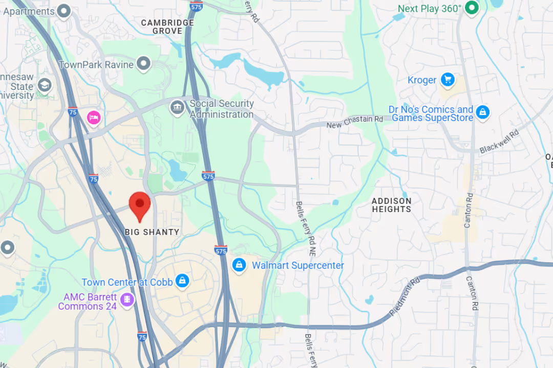
Our team delivers specialist care at convenient locations across Metro Atlanta:
Get expert tips on injury recovery, pain relief, joint health, and movement strategies—straight from our Fellowship-Trained team.