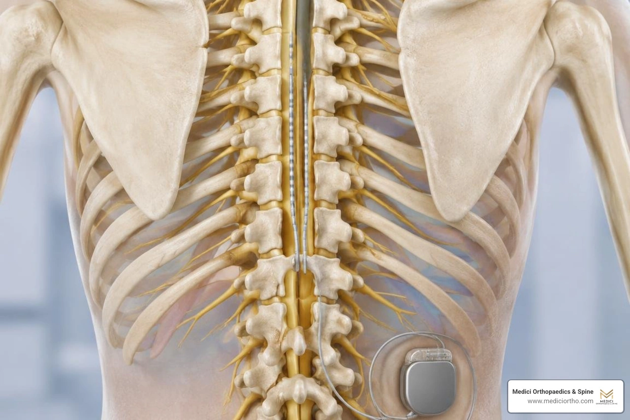
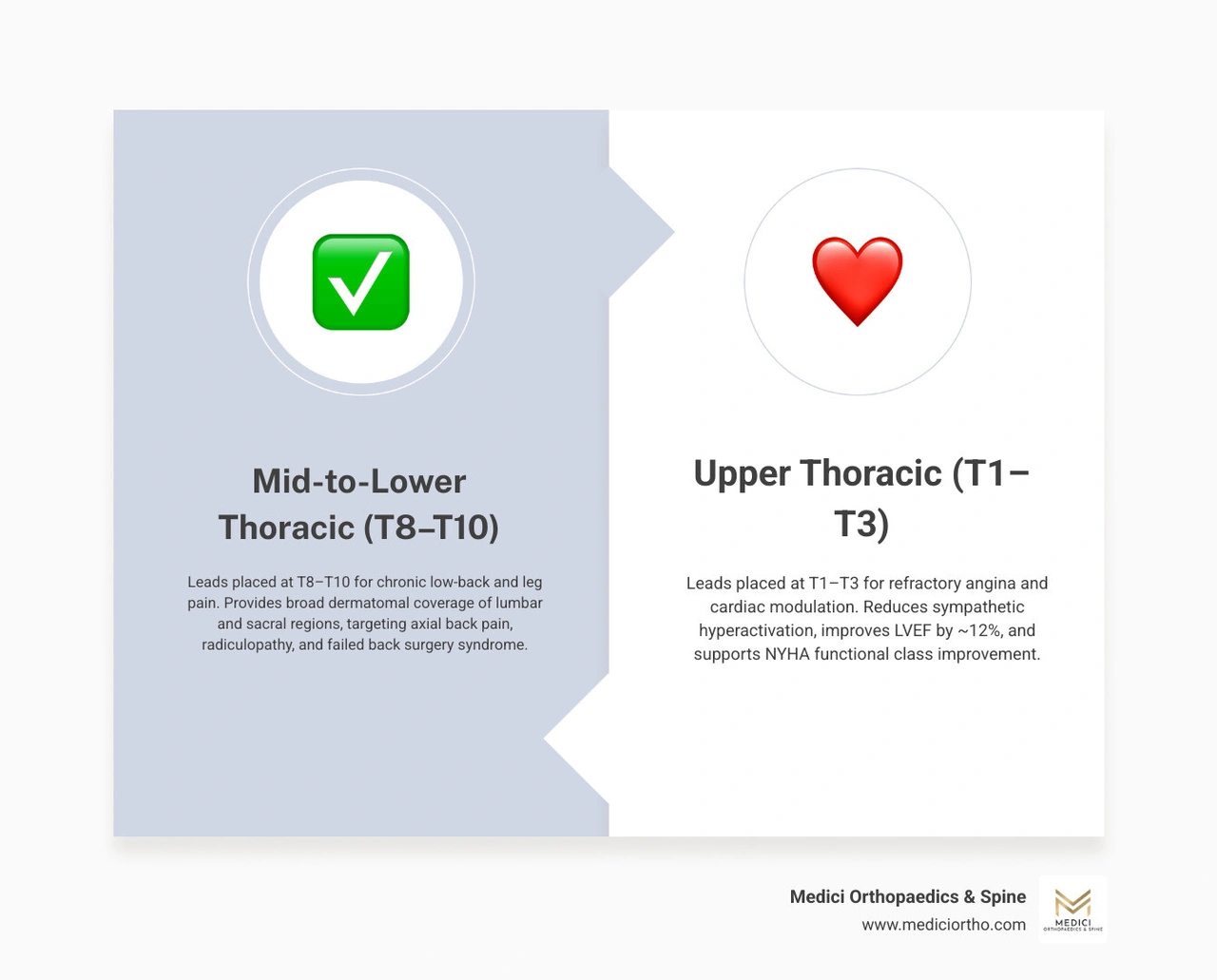
For chronic low-back and leg pain, leads are often placed near T8-T10, in the middle to lower thoracic spine. The best level depends on where you feel pain and how well stimulation covers it during a trial. Upper thoracic levels, such as T1-T3, are used for different goals, including treatment of refractory angina in select patients.
A permanent thoracic spinal cord stimulator places leads in the epidural space, just outside the spinal cord, and a pulse generator under the skin. It sends electrical pulses that change how pain signals are processed. Placement is usually considered only after other treatments have not helped enough and a trial shows meaningful relief.
I am Sonny Dosanjh, M.D., a board-certified physical medicine and rehabilitation physician with fellowship training in pain management. At Medici Orthopaedics & Spine, I help patients with chronic spine and nerve pain assess options such as a thoracic spinal cord stimulator, starting with the source of their pain and the right lead placement for their goals.
Chronic neuropathic pain can feel like an alarm system stuck in overdrive. When traditional treatments fail to quiet these rogue signals, neuromodulation offers a proven way to recalibrate the system. Understanding who qualifies for a spinal cord stimulator begins with examining how electrical pulses interrupt pain pathways before they ever reach the conscious brain.
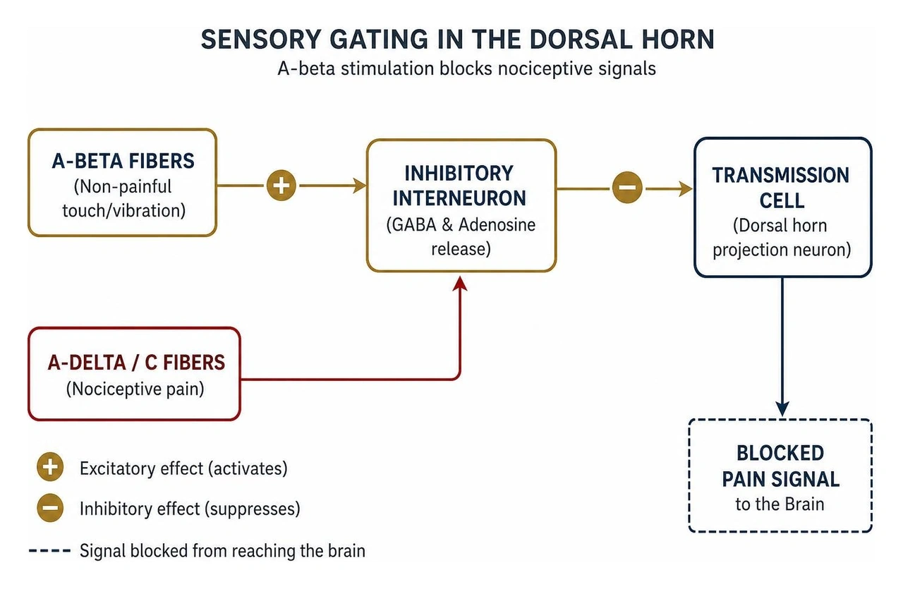
Neuromodulation relies heavily on the foundational gate control theory of pain. By introducing controlled electrical fields into the posterior epidural space, the device stimulates large-diameter A-beta sensory nerve fibers. These fibers activate local inhibitory interneurons within the dorsal horn of the spinal cord, effectively "closing the gate" against incoming nociceptive signals transmitted by smaller A-delta and C fibers (NBK553154). In addition to physical sensory gating, electrical fields trigger a biochemical cascade, promoting the local release of inhibitory neurotransmitters such as gamma-aminobutyric acid (GABA) and adenosine, which dampens hyperexcitable pain pathways (NBK553154). Comprehensive clinical summaries, such as the StatPearls review of spinal cord stimulation, highlight that this therapy consistently achieves 50% to 70% pain relief across complex neuropathic conditions.
The dorsal horn serves as the primary relay station for sensory processing. When chronic nerve injury occurs, the dorsal horn becomes sensitized, amplifying mild inputs into severe discomfort. When patients begin evaluating nerve pain stimulator options, we look closely at how the device interacts with both local spinal circuits and descending pain-modulating pathways from the brainstem.
Beyond local spinal gating, dorsal column stimulation initiates ascending signals that influence central pain processing structures, including the thalamus, anterior cingulate cortex, and somatosensory cortex. Concurrently, it enhances descending serotonergic and noradrenergic inhibitory pathways. This dual action suppresses peripheral pain signals and reduces the emotional, centralized perception of chronic discomfort.
Spinal cord stimulation is not a first-line intervention; rather, it is a highly targeted solution for intractable, medically refractory pain syndromes. At Medici Orthopaedics & Spine, we frequently employ neuromodulation for failed back surgery (also referred to as post-laminectomy syndrome), where patients continue to experience debilitating axial back and radicular leg pain despite anatomically successful surgical decompression or fusion.
Additional clinical indications include:
Placing stimulator leads at the correct thoracic vertebral level is essential for targeting specific painful areas. Because the spinal cord segments do not align perfectly with the bony vertebral bodies—the spinal cord terminates around the L1-L2 vertebral junction (the conus medullaris)—the leads must be positioned several vertebral segments above the affected dermatomes to capture the corresponding ascending dorsal column fibers.
During placement, we use real-time fluoroscopic guidance (live X-ray) to navigate the epidural space via a shallow paramedian approach. Precise midline positioning is essential; even a fraction of a millimeter lateral deviation can dramatically alter stimulation thresholds, causing unwanted unilateral stimulation or motor recruitment of the intercostal muscles.
Upper thoracic stimulation (T1–T3) targets dorsal column fibers and cardiac sympathetic afferents/efferents. In addition to treating intractable angina by reducing myocardial oxygen demand and suppressing ischemic pain perception, high thoracic neuromodulation has emerged as a groundbreaking restorative approach for cardiac dysfunction.
The landmark SCS HEART study evaluated the effects of T1–T3 stimulation in patients with severe heart failure (NYHA class III with left ventricular ejection fraction, or LVEF, between 20% and 35%). After 6 months of continuous thoracic stimulation:
By modulating excessive sympathetic drive to the heart without triggering reflex bradycardia or hypotension, upper thoracic stimulation balances autonomic tone, reduces arrhythmogenic potential, and supports myocardial mechanics.
For the vast majority of our spine and nerve patients seeking advanced stimulation therapy in Buckhead, the target zone sits within the T8 to T10 vertebral interspaces. This mid-to-lower thoracic window directly overlays the dorsal column pathways carrying sensory information from the lower extremities, buttocks, and lumbar spine.
Targeting the T9–T10 midline typically provides robust coverage across the L4, L5, and S1 dermatomes, relieving sciatica and foot pain. For axial low back pain, leads are often positioned slightly higher, across the T8–T9 region, where dorsal column fibers representing the low back dermatomes are concentrated along the physiologic midline.
Selecting the appropriate hardware configuration is just as vital as identifying the correct anatomical level. Spinal cord stimulation hardware has evolved considerably, transitioning from simple single-lead setups to sophisticated, multi-column arrays driven by advanced bioelectric waveforms.
Traditional systems relied exclusively on tonic stimulation, which generates a tingling sensation (paresthesia) to override pain signals. Today, modern neuromodulation encompasses sub-perception modalities that eliminate paresthesias altogether:
The decision between percutaneous cylindrical leads and surgically placed paddle leads depends on individual spinal anatomy, prior surgeries, canal dimensions, and long-term therapeutic goals.
Cylindrical leads are well-suited for percutaneous trials and patients with virgin thoracic spines who prefer a minimally invasive approach. Conversely, surgical paddle leads provide directional stimulation, shielding the current from anterior non-target tissues and directing it squarely into the dorsal columns. However, because paddle leads occupy more physical volume in the dorsal epidural space, careful consideration must be given to baseline canal diameter to prevent mechanical mass effect.
A major advantage of spinal cord stimulation is the two-step evaluation process: patients complete a temporary trial to confirm pain relief before moving forward with permanent implantation.
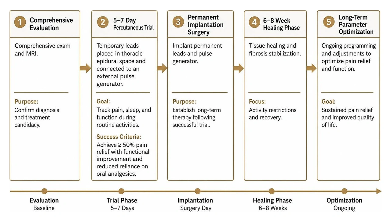
The trial phase represents an indispensable testing period. Understanding what to expect after spinal cord stimulator surgery begins by navigating the spinal stimulator trial process. Under local anesthesia and light sedation, two thin cylindrical leads are guided into the thoracic epidural space via Tuohy needles. The external ends are connected to an external pulse generator worn on a belt.
Over the course of 5 to 7 days, the patient tracks their daily pain levels, sleep quality, and physical functional capacity during routine activities. If the trial achieves at least a 50% reduction in baseline pain scores, accompanied by demonstrable functional improvements and reduced reliance on oral analgesic medications, the patient is cleared for permanent implantation.
Permanent implantation is completed as an outpatient procedure in our specialized surgical suites, providing accessible care for patients seeking outpatient spine procedures in Snellville or surgical care in Marietta.
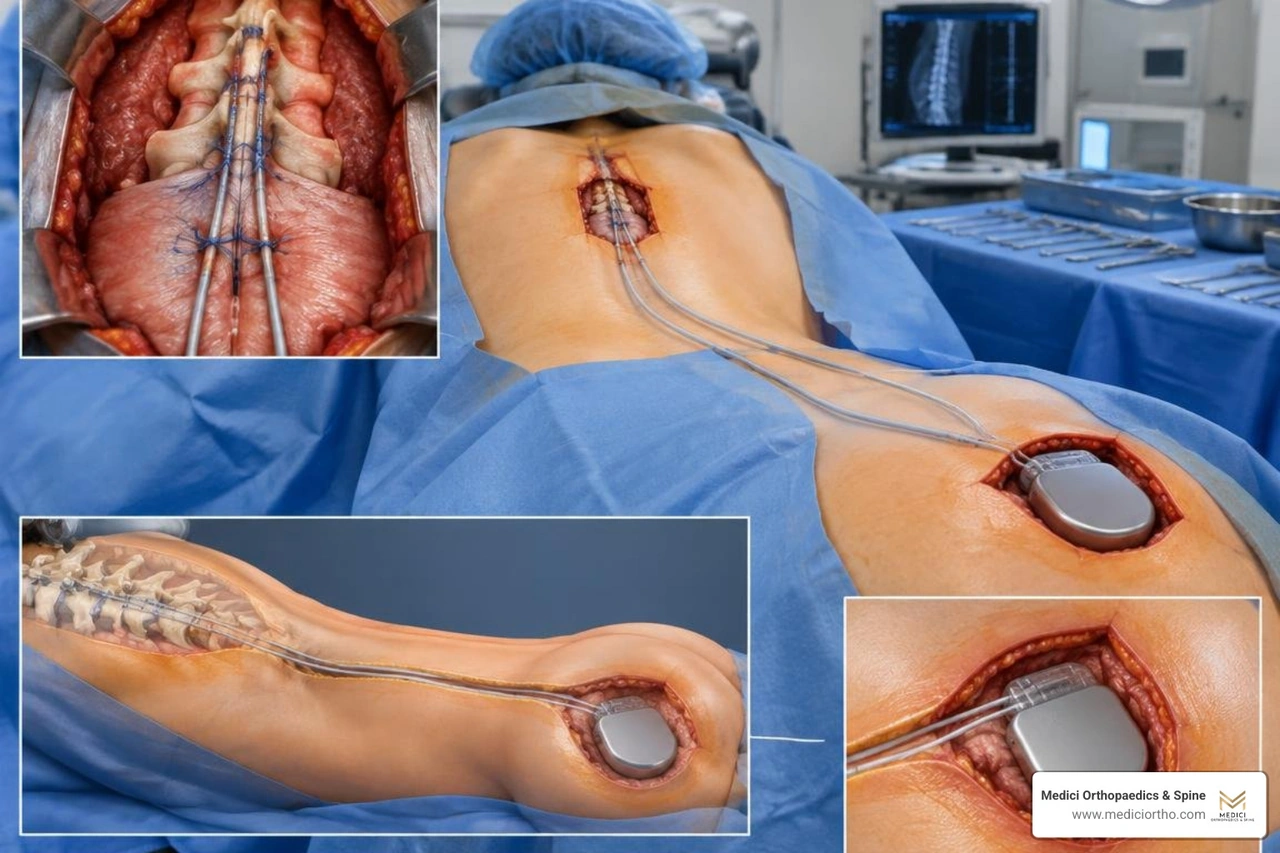
During permanent surgery:
Following permanent implantation, patients settle into stable programming parameters. However, sudden or unexpected changes in stimulation perception can serve as early diagnostic indicators of evolving spinal pathology.
In a notable clinical case, a 66-year-old patient with an established thoracic paddle lead developed new-onset lower-extremity pain and paresthesias distinct from her baseline chronic discomfort (Behzadi et al., 2021). Paradoxically, turning the stimulator's amplitude down provided immediate relief from the new symptoms, while turning the unit off eliminated them entirely (Behzadi et al., 2021).
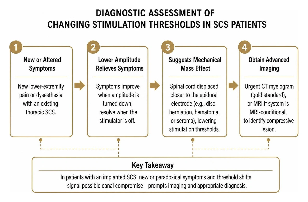
This paradoxical phenomenon occurs because a new space-occupying lesion—such as an acute thoracic disc herniation, hematoma, or seroma—displaces the spinal cord posteriorly against the fixed epidural paddle lead (Behzadi et al., 2021). The shortened distance between the electrode contacts and dorsal column fibers substantially lowers electrical impedance and perception thresholds, causing low-intensity pulses to trigger overstimulation or uncomfortable dysesthesias (Behzadi et al., 2021).
When patients with an existing stimulator present with altered paresthesias or sudden threshold shifts:
The primary red flag is the sudden onset of new, sharp, or radiating lower-extremity pain that feels distinctly different in character or location from your original baseline condition (Behzadi et al., 2021). If your standard stimulation settings suddenly feel uncomfortable, or if symptoms resolve when you significantly turn down the device amplitude, this indicates that the physical distance between the leads and your spinal cord has changed (Behzadi et al., 2021). This mechanical mass effect warrants an urgent clinical evaluation and diagnostic CT myelography or MRI to rule out an acute thoracic disc herniation, epidural hematoma, or seroma (Behzadi et al., 2021).
Upper thoracic neuromodulation (specifically targeting the T1–T3 spinal segments) directly modulates the cardiac nervous system and sympathetic outflow tracts. In clinical trials such as the SCS HEART study, modulating these autonomic pathways reduced cardiac stress and improved left ventricular ejection fraction by an average of 12% over six months. It also decreased left ventricular end-systolic volume and improved functional NYHA class status without causing adverse device-device interactions in patients with existing cardioverter-defibrillators.
The initial surgical incisions generally heal within 10 to 14 days, during which you must keep the dressings clean and dry. However, the internal healing phase lasts approximately 6 to 8 weeks. During this critical window, your body forms a protective layer of fibrous connective tissue around the epidural leads, anchoring them securely in place. You will need to avoid heavy lifting (over 10 pounds), aggressive torso twisting, bending, or reaching overhead to minimize the risk of early lead migration. Most patients resume light, non-strenuous daily activities within 2 to 3 weeks.
Living with intractable chronic pain can diminish your independence, limit your career, and disrupt your daily life. At Medici Orthopaedics & Spine, we believe you should not have to depend indefinitely on high-dose medications or undergo repeated, invasive open spine surgeries to find meaningful relief.
Our practice is dedicated to helping you restore your quality of life through the most effective, least invasive, and least drug-dependent therapies available. Because individual pain conditions are complex, we know that treatments work best when integrated together. Physical therapy, targeted injections, regenerative medicine, and advanced spinal cord neuromodulation work synergistically to help you regain functional freedom.
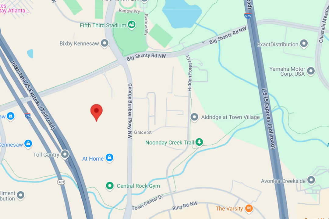
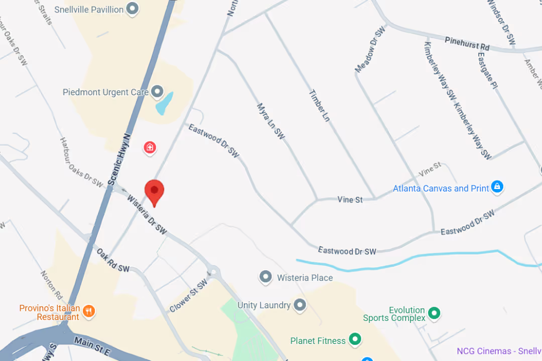
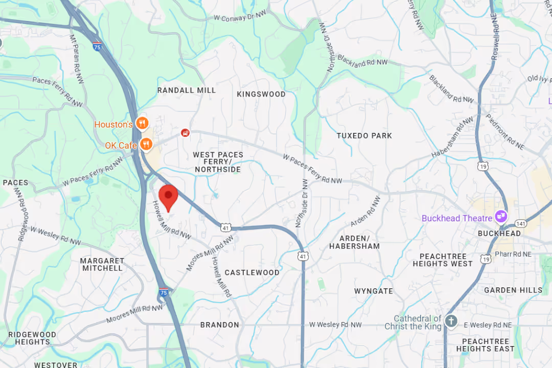
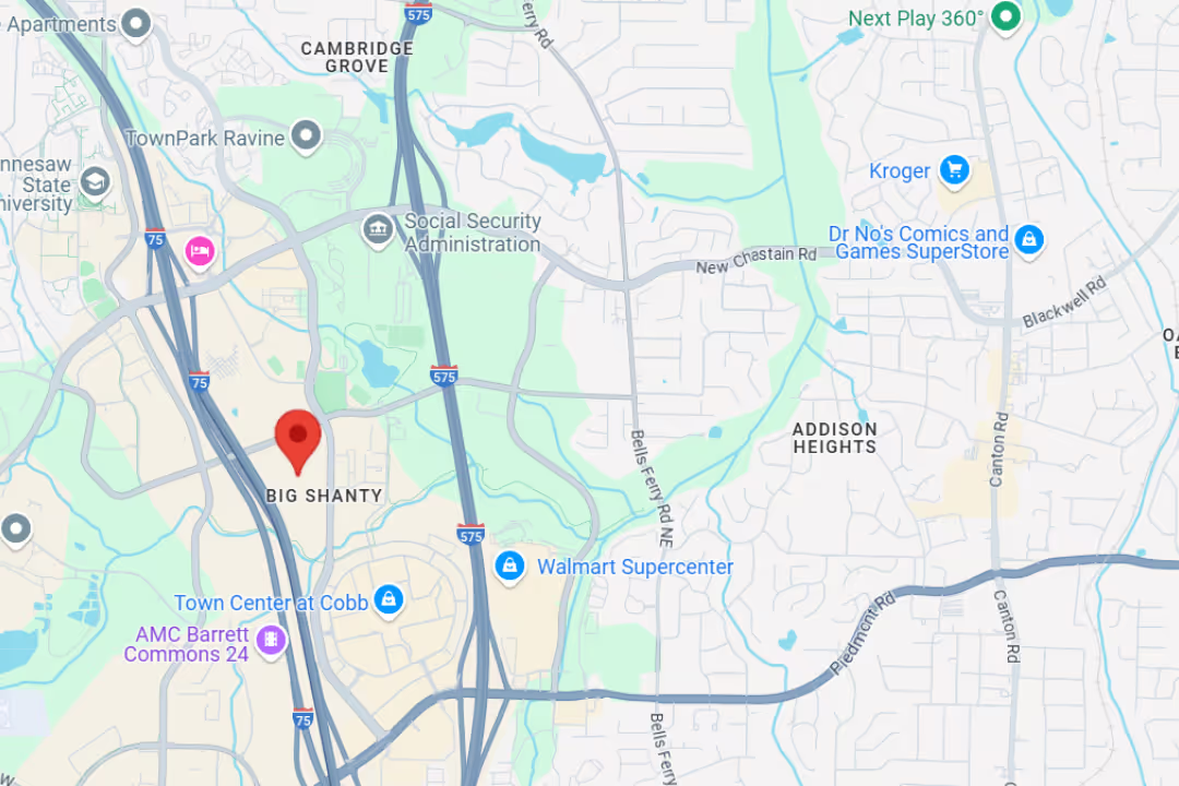
If chronic axial back pain, post-surgical neuropathy, or severe nerve injuries are limiting your daily life, take the next step. Contact our team to schedule an evaluation for advanced spinal cord stimulation at our state-of-the-art facilities in Snellville, Marietta, Kennesaw, or Buckhead, and let us design a personalized recovery plan tailored to your lifestyle.
NBK553154.Behzadi F, Telemi E, Mansour TR, Zervos TM, Abdulhak MM, Air EL. "Acute thoracic disc heralded by change in spinal cord stimulation pattern: illustrative case.." Journal of neurosurgery. Case lessons, 2021. PMCID PMC9435567.White T, Justiz R, Almonte W, Micovic V, Shah B, Anderson E, Kapural L, Cordner H, El-Naggar A, Fishman M, Eshraghi Y, Kim P, Abd-Elsayed A, Chakravarthy K, Millet Y, Sanapati M, Harrison N, Goff B, Gupta M, Grewal P, Wilkinson M, Bundschu R, Will A, Satija P, Li S, Dulebohn S, Broadnax J, Gekht G, Wu K, Falowski S, Park W, Cedeno DL, Vallejo R. "Twelve-month results from a randomized controlled trial comparing differential target multiplexed spinal cord stimulation and conventional spinal cord stimulation in subjects with chronic refractory axial low back pain not eligible for spine surgery.." North American Spine Society journal, 2024. PMCID PMC11369449.
At Medici, you’re more than your MRI.
We take time to hear your story, understand your pain, and create a plan that actually works for you.

Our team delivers specialist care at convenient locations across Metro Atlanta:
Get expert tips on injury recovery, pain relief, joint health, and movement strategies—straight from our Fellowship-Trained team.